[ad_1]
LyOK-432 had a stronger selling impact on DC maturation
As proven in Extra file Supplies (Extra file 1: Determine S1), the speed of mature DCs (CD80+CD86+ DC) was considerably larger within the lyOK432 group than within the OK-432 and lipopolysaccharide (LPS) teams (p < 0.001 and p < 0.001, respectively). Moreover, DCs within the lyOK-432 group had the best expression ranges of IL-6 and IL-12 in contrast with the opposite three teams (p < 0.001). These outcomes indicated that lyOK-432 promoted DC maturation extra effectively than non-lysed OK-432.
Preparation and characterization of ROD
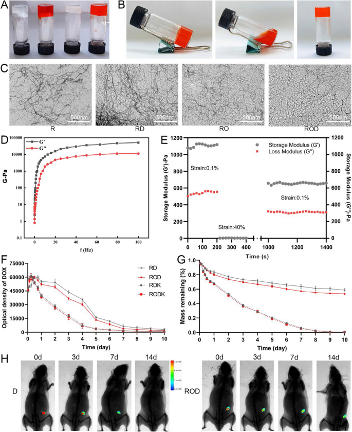
The synthesis of ROD, RO, RD, and R was described within the Supplies and Strategies part. The RADA16-I peptide hydrogel was reported platform for loading a broad vary of hydrophilic and hydrophobic parts [21]. The 4 sorts of peptide hydrogels on this examine had been additionally efficiently shaped (Fig. 1A). To find out the secure loading capability of ROD, we first modified the pH situation and outcomes confirmed that the ROD peptide gelated at pH of 4.5–7.5, however didn’t type at 8.5 (Extra file 1: Determine S2), indicating the gelation capability beneath physiological situations and in tumor tissue. As well as, after repeated vigorous shaking, the ROD hydrogel turned an injectable answer, indicating its thixotropic character (Fig. 1B).
Preparation and characterization of ROD hydrogel. A R, RD, RO, and ROD hydrogel gelated for 7 days. B Thixotropic property of the ROD hydrogel. C TEM photos of R, RD, RO, and ROD hydrogel, exhibiting that these hydrogels self-assembled into networks of interwoven nanofibers. D Frequency sweep rheological evaluation of the ROD hydrogel, suggesting the formation of a secure hydrogel. E Step-strain time-dependent rheological evaluation of the ROD hydrogel, indicating ROD had rheological nature of for injection. Measurements had been carried out at a hard and fast angular frequency of 1 rad/s. F The discharge profiles of RD and ROD hydrogel-loaded DOX within the presence or absence of proteinase Ok. G The measurement of the load of RD and ROD hydrogels within the presence or absence of proteinase Ok. H The discharge profile of DOX in ROD hydrogel and DOX answer after intratumor injection within the subcutaneous Hep1-6 tumor (n = 3), suggesting controlled-release nature of ROD hydrogel. ROD, RADA16-I peptide hydrogel loaded with lyOK-432 and doxorubicin; RO, RADA16-I peptide hydrogel loaded with lyOK-432; RD, RADA16-I peptide hydrogel loaded with doxorubicin; R, RADA16-I peptide hydrogel; DOX D doxorubicin, TEM transmission electron microscopy
TEM was used to guage the dimensions and morphology of the ROD hydrogel. As proven in Fig. 1C, the ROD hydrogel self-assembled into networks of interwoven nanofibers. The rheological traits of the ROD hydrogel was evaluated by way of the storage modulus (G’) and loss modulus (G’’). Determine 1D exhibits that G’ and G’’ had been much less depending on the frequency (0.1–100 rad/s) because the pressure fixed elevated, which advised the formation of a secure hydrogel. As well as, a time-dependent step-strain rheological experiment was performed to guage the injectability of the ROD hydrogel. Throughout this course of, a low fixed pressure of 0.1%, a better pressure of 40%, and a restoration pressure of 0.1% had been examined and recorded. Determine 1E exhibits that the ROD hydrogel just about restored its unique power at 1000 s after withdrawal of the bigger pressure, which indicated that the ROD possessed rheological nature for injection.
Drug launch in vitro and in vivo
The discharge of DOX from the ROD hydrogel, with or with out proteinase Ok, was measured in vitro utilizing a fluorescence spectrophotometer. As proven in Fig. 1F, the optical density elevated within the first 18 h, however step by step decreased thereafter within the 4 teams and was virtually undetectable 10 days later. As well as, the optical density of DOX dropped steeply within the presence of proteinase Ok, suggesting an elevated launch of DOX from the hydrogel. Alternatively, the remaining peptide spine of the ROD hydrogel was weighed. The outcomes (Fig. 1G) confirmed that 53.5% of the ROD hydrogel remained after 10 days whereas it was utterly digested with proteinase Ok, which advised a superb biodegradability of this hydrogel.
A subcutaneous Hep1-6 tumor mannequin was established to detect drug launch in vivo (Fig. 1H). After the tumor quantity reached ∼100 mm3, the fluorescence depth was excessive after intratumoral injection of 100 μL ROD or DOX on day 0. Nevertheless, the fluorescence sign within the DOX group turned weaker 3 and seven days later, and was nearly undetectable on the 14th day. In distinction, the fluorescence depth after ROD injection was stronger on days 3 and seven, and the sign might nonetheless be detected 14 days later. Moreover, the fluorescence sign within the RD group was akin to that of the ROD group, indicating that the incorporation of lyOK-432 didn’t alter the discharge profile of DOX within the hydrogel (Extra file 1: Determine S3). Collectively, these outcomes advised that ROD had a controlled-release nature.
Antitumor impact of ROD
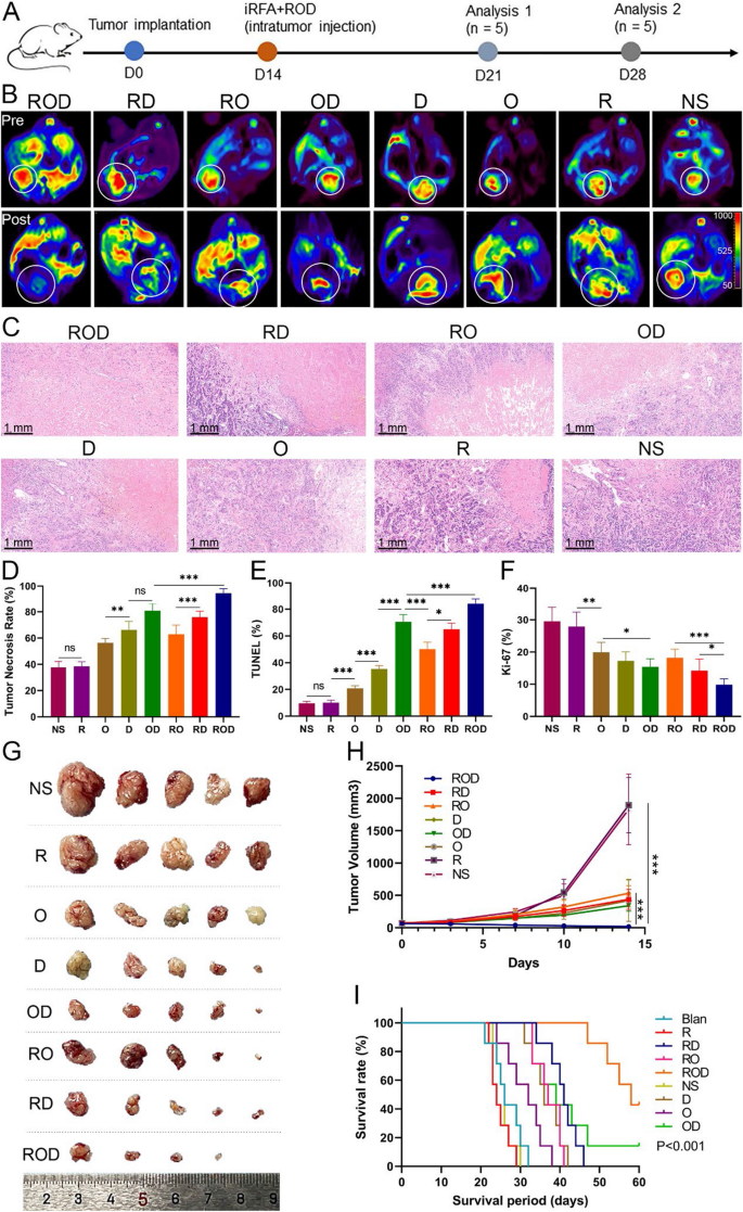
To discover the anticancer impact of ROD hydrogel, a mouse mannequin of orthotopic HCC was established. After iRFA, the residual viable tumor tissues in numerous teams had been intratumorally injected with ROD, RO, RD, OD, O, D, R, and NS, respectively (Extra file 1: Determine S4). Tumor response was evaluated 14 days after therapy. As proven by the DWI photos in Fig. 2B, the residual tumor tissues within the ROD group had been hardly ever seen, whereas the alerts of viable tumors had been conspicuously stronger in different teams, particularly within the NS and clean teams. As well as, the tumor necrosis fee was calculated based on HE staining, as beforehand described [23]. Outcomes confirmed that the necrosis fee was 94.6% ± 1.5% within the ROD group, which was considerably larger than within the different 7 teams (75.9% ± 2.0% within the RD group, 62.9% ± 3.2% within the RO group, 80.8% ± 2.5% within the OD group, 66.3% ± 2.9% within the D group, 56.5% ± 1.4% within the O group, 38.4% ± 1.6% within the R group, and 37.6% ± 2.0% within the NS group, respectively; p < 0.001, Fig. 2C, D). TUNEL yield the best fee of constructive staining, whereas Ki-67 produced the bottom fee within the ROD group (p < 0.001, Fig. 2E, F, and Extra file 1: Determine S5), the findings being in step with the outcomes of the DWI imaging and HE staining. The tumor quantity 14 days after therapy was additionally considerably smaller within the ROD group than within the different teams (p < 0.05, Fig. 2G, H).
Antitumor impact of ROD hydrogel (n = 5). A Therapy schedule of mice bearing HCC. Evaluation 1 refers back to the analysis of antitumor immunity, and evaluation 2 refers back to the analysis of tumor therapy efficacy. B DWI photos exhibiting the residual tumor tissues 14 days after therapies. C Consultant HE-stained tumor sections (×10 magnification). D–F Quantitative evaluation of tumor necrosis, apoptosis, and proliferation on the idea of HE staining, TUNEL assay, and Ki-67 staining. These outcomes indicated the optimum antitumor impact was achieved in ROD group. G {Photograph} of dissected tumor samples. H Measurement of tumor volumes on the indicated instances. Tumor development was considerably inhibited in ROD group. I Kaplan–Meier survival curves present that the survival time of mice was considerably longer within the ROD group than in different teams (n = 7). DWI diffusion weighted imaging, NS regular saline; O, OK-432. Ns not vital; *p < 0.05; **p < 0.01; ***p < 0.001
Moreover, survival evaluation was performed for the iRFA-treated mice within the 8 teams (Fig. 2I). Three mice (37.5%) within the ROD group survived for greater than 60 days, which was considerably longer than the median survival time within the RD group (41 days), RO group (36 days), OD group (39 days), D group (36 days), O group (32 days), R group (24 days), NS group (26 days), and clean teams (26 days) (p < 0.001). Collectively, these outcomes demonstrated that ROD therapy achieved a potent synergistic antitumor impact in opposition to liver most cancers after iRFA remedy.
ROD enhanced in vivo antitumor immunity
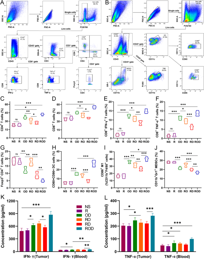
To determine the immunological elements that impression therapeutic efficacy after iRFA, tumor-infiltrating lymphocytes (TILs) had been evaluated after therapies, since earlier research indicated that DOX and OK-432 might set off antitumor immunity [12, 19]. The tumor tissues had been harvested at 7 days after therapies, and the immune cells had been counted by circulation cytometry. As offered in Fig. 3A–J, the outcomes revealed that the ROD group had the best ranges of CD4+, CD8+ T cells and M1 polarized tumor-associated macrophages (M1-TAMs), whereas the R and NS teams had the decrease counts (p < 0.001). The outcomes of TAM and M2-TAM had been proven in Extra file 1: Determine S6. Furthermore, the proportions of CD4+ T cells, CD8+ T cells, and M1-TAM had been additionally considerably larger within the RO and RD teams than within the R and NS teams (p < 0.001). In distinction, the proportion of Tregs and myeloid-derived suppressor cells (MDSCs) within the ROD group was considerably decreased in contrast with different teams (p < 0.01, Fig. 3G, J), and was considerably elevated within the R and NS teams when put next with the clean controls (p < 0.001). Moreover, the degrees of IFN-γ and TNF-α in tumor tissues and blood had been decided by flowcytometry and ELISA (Fig. 3E, F, Ok, L). The outcomes confirmed that these cytokines had been all remarkably elevated within the ROD group (p < 0.001), however had been mildly elevated and considerably larger within the RO, RD, and OD teams in contrast with the NS and clean controls (p < 0.05). Immunohistochemical staining of TILs additional demonstrated the findings (Extra file 1: Determine S7). Nevertheless, the clearance of CD4+ T cells, CD8+ T cells, or each with antibodies remarkably impaired the antitumor efficacy of the ROD hydrogel (knowledge not proven). Taken collectively, these outcomes demonstrated that the ROD hydrogel effectively improved the infiltration, accumulation, and performance of antitumor immune cells.
Enhanced antitumor immunity after ROD remedy (n = 5). A, B Consultant dot plots of the morphological traits (SSC vs FSC) of tumors subjected to ROD. Schematic illustration of gating: Tumor-infiltrating lymphocytes had been stained with corresponding antibodies for circulation cytometric evaluation, together with CD4+ T, CD8+ T, Treg cells, DCs, TAM, and MDSCs. C–J Quantitative evaluation of the odds of CD4+ T, CD8+ T, Foxp3+CD4+ T, DCs, M1, and MDSCs in tumors 7 days after therapies. Ok, L Expressions of IFN-γ and TNF-α in tumor tissues and blood. DC dendritic cell, TAM tumor-associated macrophage, MDSCs myeloid-derived suppressor cell, IFN interferon, TNF tumor necrosis issue. Ns not vital; *p < 0.05; **p < 0.01; ***p < 0.001
Improved antitumor immunity by way of enhanced cGAS/STING pathway in DCs
To additional perceive the mechanism of enhanced antitumor immune responses, DCs had been measured by circulation cytometry, which is the potent antigen presenting cells for activating the antitumor immune cells (Fig. 3A, H). The ROD group displayed the best stage of mature CD80+CD86+ DCs (mDC) in contrast with the opposite teams (p < 0.001). The proportion of mDCs within the RO, RD, and OD teams was considerably larger than within the NS group (p < 0.05). Immunohistochemical staining additionally confirmed these outcomes (Extra file 1: Determine S7). Nevertheless, blocking the DCs with antibody within the ROD group decrease the tumor necrosis fee, which akin to that of the RD and OD group (knowledge not proven). Collectively, these outcomes indicated that ROD, RO, RD, and OD therapy promoted DC maturation, adopted by enhanced antigen presentation and immune cell recruitment within the residual liver most cancers tissues 7 days after iRFA.
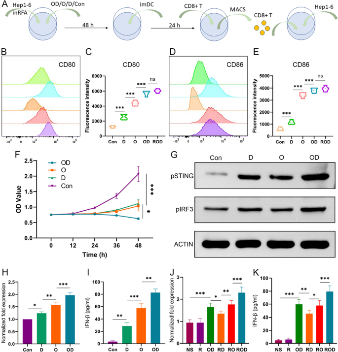
Earlier research have indicated that OK-432, as a international antigen, might straight recruit DCs and promote their maturation, whereas DOX in all probability activate DCs alongside different pathways [12, 20]. Based mostly on these hypotheses, we investigated the synergistic impact of ROD by analyzing the cGAS/STING/IFN-I signaling pathway in DCs each in vitro and in vivo. In in vitro experiments, Hep1-6 cells obtained iRFA administration and ROD, OD, O, D, or left untreated (clean controls), and co-cultured with imDCs after 48 h (Fig. 4A). The floor markers of CD80+CD86+ mDCs had been decided by circulation cytometry. As proven in Fig. 4B–E, the proportion of mDC considerably elevated after ROD and OD therapy (p < 0.01). As well as, hep1-6 cells had been cocultured with CD8+ T cells after co-incubated with hep 1–6 specialised mature DCs. The outcomes of CCK-8 confirmed a suppressed viability of Hep1-6 cells within the OD, O, and D teams in contrast with the management group (Fig. 4F), indicating an enhanced antigen presentation between DCs and CD8+ T cells.
LyOK-432 together with DOX activated cGAS/STING pathway in DCs (n = 3). A In vitro illustration of examine schedule for co-culture of Hep 1–6 cells, imDC, and CD8+ T cells after administration of iRFA and OD, O, D, or management. (B–E) Quantitative evaluation of the floor makers of mature DCs (CD80+ CD86+ DC). The expression stage of CD80 and CD86 was highest in ROD/OD group. F The cytotoxicity of CD8+ T cells induced in vitro in opposition to Hep1-6 cells within the 4 teams as revealed by CCK-8 assay. The most cancers cells had been considerably suppressed after OD administration. G Western blotting confirmed that the protein expression of phosphorylated STING and IRF3 was larger in OD group than within the different teams. H, I The IFN-β expression measured by qPCR and ELISA after completely different in vitro therapies. J, Ok The IFN-β expression in tumor tissues measured by qPCR and ELISA after completely different in vivo therapies (n = 5). cGAS, cyclic guanosine phospho-adenosine synthase; STING, stimulator of interferon genes; MACS, magnetic cell sorting. *p < 0.05; **p < 0.01; ***p < 0.001
To verify the activation of the cGAS/STING/IFN-I signaling pathway, the protein ranges of phosphorylated STING (pSTING) and phosphorylated IRF3 (pIRF3, a downstream marker) had been measured by Western blotting in in vitro experiment. Moreover, IFN-β expression was decided by qPCR and ELISA. As proven in Fig. 4G, the expressions of pSTING and pIRF3 within the OD group had been larger than within the different group, and the degrees within the O and D teams had been additionally larger than within the management teams. Moreover, the expression ranges of IFN-β in these teams had been in step with these of the pSTING and pIRF3 proteins. Amongst them, the OD group had the best expression stage (Fig. 4H, I) (p < 0.01). Furthermore, after inhibiting the expression of STING by a particular inhibitor (c-176), IFN-β ranges didn’t improve clearly in contrast with the management group (knowledge not proven). Alternatively, the expressions of pSTING, pIRF3, and IFN-β had been evaluated in Hep1-6 cells to rule out different potential secretion sources. As anticipated, they had been expressed at a low stage akin to the management group (knowledge not proven).
The expression of IFN-β was additionally decided in tumor tissues in vivo (Fig. 4J, Ok). As anticipated, IFN-β stage was considerably up-regulated within the ROD group in contrast with the opposite teams (p < 0.05). Taken collectively, these outcomes indicated that the synergistic impact of ROD hydrogel promoted the recruitment and maturation of imDCs, activated the cGAS/STING/IFN-I pathway, enhanced the antitumor immunity, and thus improved the therapy efficacy in opposition to residual liver most cancers after iRFA remedy.
Furthermore, the expressions of IFN-β, pSTING and pIRF3 proteins in DCs had been additionally in contrast among the many teams of lyOK-432, OK-432, LPS, and controls (Extra file 1: Determine S1). The outcomes confirmed that the mRNA and cytokine ranges of IFN-β within the lyOK-432 group had been considerably larger than within the OK-432 group (p < 0.05), and the protein ranges of pSTING and pIRF3 had been additionally clearly larger within the lyOK-432 group when put next with different teams. These outcomes indicated that lyOK-432 additionally promoted the activation of the cGAS/STING/IFN-I pathway extra successfully than non-lysed OK-432.
Immune reminiscence results of ROD therapy
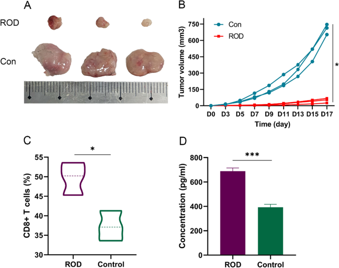
Because the ROD hydrogel therapy offered a potent tumor-inhibitory impact and triggered antitumor immunity, we performed a re-challenge check to analyze whether or not an immune reminiscence impact was generated. Mice that survived within the survival experiment (n = 3 within the ROD group) and clean mice (with out pre-tumor-bearing, n = 3) had been examined. Hep1-6 cells (1 × 10⁶) had been subcutaneously injected, and the tumor quantity was recorded. As proven in Fig. 5A, B, tumor progress was considerably suppressed within the ROD group (p = 0.01), which demonstrated {that a} sturdy immune reminiscence impact was generated in these surviving mice.
Immune reminiscence impact within the mice that survived after ROD remedy (n = 3). A {Photograph} of dissected tumor samples within the ROD group and clean management group. B Measurement of tumor volumes on the indicated instances. The subcutaneous tumors had been considerably inhibited in ROD group. C, D Quantitative evaluation of CD8+ T cells and IFN-γ within the subcutaneous tumors after therapy s, which was considerably larger in ROD group. *p < 0.05; ***p < 0.001
CD8+ T cells and IFN-γ had been measured within the tumor tissues of the 2 teams, and the outcomes advised that the proportion of CD8+ T cells and the expression of IFN-γ had been each considerably larger than within the management group (p = 0.02 and < 0.001, respectively, Fig. 5C, D).
Security analysis
Liver and renal operate exams had been carried out in every group on the indicated time factors (Fig. 6). The outcomes advised that these therapies had little impact on mice liver and renal features 7 days later. HE staining revealed regular morphologies of main organs, together with the guts, liver, spleen, lung, and kidney. The physique weight of mice after therapy skilled no vital change in contrast with the management group (p = 0.34). These outcomes indicated that the ROD hydrogel is secure as a therapy for the residual most cancers after iRFA of HCC.
Security evaluation of ROD remedy (n = 5). A Liver and renal operate had been examined 7 days after therapies. B HE staining of the guts, liver, spleen, lung, and kidney samples (×20 magnification). C Mice physique weight on the indicated instances after completely different therapies. These outcomes offered that ROD therapy don’t have any further injury to mice. BUN blood urea nitrogen, Cre creatinine, AST aspartate aminotransferase, ALT alanine aminotransferase. Ns not vital
[ad_2]

